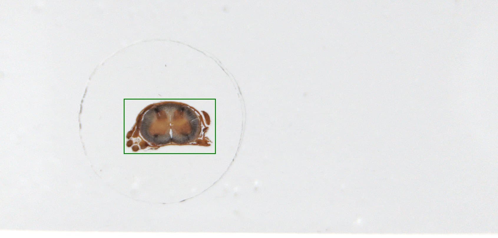
Spinal Cord
Illuminator: 35.0
Brightfield
Brightfield
Fields: 323 (17×19) HD
36.956mm² (7.878mm × 4.691mm)
36.956mm² (7.878mm × 4.691mm)
Key Commands
- ↑ or W
- Move Viewport Up
- ↓ or S
- Move Viewport Down
- ← or A
- Move Viewport Left
- → or D
- Move Viewport Right
- 0 or *
- Move Viewport Home
- [shift] W, +, =
- Zoom In
- [shift] S, -, _
- Zoom Out
Menu Buttons
- Maximize Display
- Restore Display
- Move Viewport Home
- Zoom In
- Zoom Out
- Rotate Left 90°
- Rotate Right 90°
- Filter Settings
- Toggle Scalebar
- Toggle Navigator
- Scan Information
- Help Window
- Job Name
- SpinalCord-HX20-HDX
- Scan Date
- 13-Oct-2017 14:54
- Scan Duration (hh:mm:ss)
- 00:07:51
- Process Duration (hh:mm:ss)
- 00:01:06
- Model
- uScopeHXII
- Objective
- 20x (0.65NA)
- Image Format
- HD JPEG (Q90)
- Pixel Mapping
- 0.258µm/pixel
- Focus Method
- Exhaustive Stack
- Illuminator
- 35.0
- Fields
- 323 (17 × 19)
- Region Size
- 36.956mm² (7.878mm × 4.691mm)
- Overview Image

Reset
Related Whole Slide Images
Scanned by the uScope Digital Microscope
Scanned by the uScope Digital Microscope
Contact Microscopes International or speak with your local distributor.
Copyright © Microscopes International, LLC. All rights reserved.


